Nobel Laureate who discovered the HIV presents controversial but well-documented findings that electromagnetic signals can be detected from highly diluted solutions of DNA. Dr. Mae-Wan Ho
“Luc Montagnier, the French virologist who won a Nobel prize in 2008 for linking HIV with AIDS, last week made controversial claims that highly dilute solutions of harmful viruses and bacteria emit low-frequency radio waves, allegedly from watery nanostructures formed around the pathogens. Similar claims have been made for homeopathic remedies.” New Scientist [1]
Homeopathy has been subject to periodic attacks from the mainstream medical and scientific community aided and abetted by uninformed journalist in the mainstream press eager to create a good impression with the scientific establishment.
The latest round was initiated by a damning report from the UK Parliament Science and Technology Committee released in February 2010, Evidence Check 2: Homeopathy [2], which concludes that the existing scientific literature shows no evidence that that homeopathy is efficacious beyond the placebo effect, and that “explanations for why homeopathy would work are scientifically implausible.” Therefore, the National Health Service should stop funding homeopathy and the Medicines and Healthcare products Regulatory Agency should not allow homeopathic product labels to make medical claims without evidence of efficacy.
In July, the British Medical Association passed a resolution to stop homeopathy being made available on the National Health Service (NHS), and to have all homeopathic remedies to be placed in a special area marked ‘Placebos’ in health shops and pharmacies. However, the UK government is not taking action to ban homeopathy from the NHS [3], which has funded homeopathy from its inception in 1948. So homeopathy is safe, at least for now.
The most difficult hurdle in getting general acceptance for homeopathy is without doubt the lack of an explanation, based on contemporary science, on why it would work. In my view, that is more important than getting double-blind, placebo-controlled data on efficacy. Such an explanation is beginning to emerge, and Luc Montagnier’s research team may have provided some key observations.
The Nobel Laureate has entered the fray, bravely picking up on work done by his fellow countryman, the recently deceased immunologist Jacques Benveniste, who became the centre of a major international controversy in 1988, when Benveniste and his research team published a paper in the journal Nature describing the apparent homeopathic action of very high dilutions of anti-IgE antibody on the human blood cells basophils. As condition for publishing the paper, the then journal editor John Maddox organised and subjected Benveniste and his team to a farcical and damaging public trial [4] that included illusionist and well-known sceptic James Randi and fraud expert Walter Stewart .
Montagnier’s recent work, summarily dismissed in the New Scientist [1] and elsewhere, has been published in two papers in 2009, and the evidence presented is clear and informative.
The first paper reports the capacity of some bacterial DNA sequences to induce electromagnetic waves at high dilutions in water [5], and appears to be a “resonance phenomenon” triggered by the ambient electromagnetic background of very low frequency waves. Interestingly, genomic DNA of most pathogenic bacteria contain sequences that are able to generate such signals, suggesting that highly sensitive detection system might be developed for chronic bacterial infections in human and animal diseases. The second paper follows up this suggestion, showing that it is indeed possible to detect the presence of HIV DNA even when the RNA of the virus has disappeared from the blood of people infected with HIV and undergoing antiviral therapy (see [6] Electromagnetic Signals from HIV, Prospects for a Science of Homeopathy, SiS 48).
Montagnier and his colleague Claude Lavallee initially observed that filtering a culture supernatant of human lymphocytes infected with the bacterium Mycoplasma pirum (about 300 nm in diameter) through filters with pore size of 100 nm or 20 nm gave apparently sterile fluid. However, the sterile fluid was able to regenerate the original mycoplasma when incubated with a mycoplasma-negative culture of human lymphocytes within 2 to 3 weeks. Similarly, filtering an infective fraction of HIV particles (120 nm) through 20 nm filter failed to retain the infective agent.
Furthermore, the infectious filtrate produced electromagnetic waves of low frequency in a reproducible manner after appropriate dilutions in water. They suspected a “resonance phenomenon” depending on excitation by the ambient electromagnetic fields such as the 50/60 Hz signals from the mains. The infectious signal appeared associated with “polymeric nanostructures of defined size” present in the diluted filtrate. The supernatant of uninfected eukaryotic cells used as controls did not have those infectious effects.
Given the initial clues, the researcher team set out to investigate the phenomenon more thoroughly, to characterize the electromagnetic (EM) signals and the nanostructures produced by the purified bacteria.
In addition to M. pirum, they looked at E. coli. The supernatants of deliberately infected human lymphocytes containing 106 or 107 infectious units per ml were filtered twice first through 450 nm Millipore filters to remove debris, and then 100 or 20 nm filters to remove mycoplasma cells. The filtrates were confirmed sterile by incubation for several weeks in enriched growth medium. Repeated search for traces of mycoplasma DNA by polymerase chain reactions (PCR) was also consistently negative.
However, when the filtrates were incubated for two weeks or three weeks with a culture of human activated T lymphocytes, the mycoplasma was recovered in the medium with all its original characteristics.
The filtrates were analysed just after filtration for production of EM waves of low frequency. For this purpose, a devise previously designed by Benveniste and Coll was used for the detection of signals produced (see Figure 1).
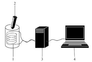
Figure 1 Detecting EM signals with Benveniste and Coll’s device
The filtrates were serially diluted 1 in 10, after each dilution, the tube is tightly stopped and strongly agitated on a Vortex apparatus for 15 seconds. This step, which is equivalent to homeopathic ‘succussion’, has been found critical for the generation of signals.
After all dilutions have been made (15 to 20), the stopped tubes were read one by one on an electromagnetic coil (copper wire on a bobbin, impedance 300 Ohms), connected to a Sound Blaster Card, itself connected to a laptop computer powered by its 12 volt battery. Each emission is recorded twice for 6 seconds, amplified 500 times and processed with different softwares to visualise the signals on the computer screen. The main harmonics of the complex signals were analysed by softwares for Fourier transformations. In each experiment, the internal noise generated by the different pieces of the reading system was first recorded (coil alone, coil with a tube filled with ordinary water). Fourier analysis shows that the noise was predominantly very low frequencies probably generated at least in part by the 50/60 Hz ambient electric current. Using the 12 volt battery to power the computer reduced the noise, but did not abolish it altogether; as the noise was found to be necessary for the induction of the resonance signals from the specific nanostructure.
When the EM signals from serial dilutions of the M. pirum filtrate were recorded, the first obvious change was an increase in the overall amplitude of the signals at certain dilutions over the background noise, and also higher frequencies. This change was abolished if the tube analysed was placed inside a box shielded with sheets of copper and mumetal, which also shields static magnetic field as well as low frequency EM fields.
Fourier analysis of the M. pirum signals confirmed a shift towards higher frequencies close to 1 000 Hz and multiples thereof. The profiles were identical and highly reproducible for all the dilutions showing an increase in amplitude.
The first low dilutions were usually negative, showing the background noise only, positive signals were typically obtained at dilutions ranging from 10-5 to 10-8 up to 10-12, at which the signal was greatest before it became negative at 10-13.
The positive dilutions varied according to the type of filtration; the 20 nm filtrate being generally positive at dilutions higher than those of the 100 nm. The original unfiltered suspension was negative at all dilutions, a phenomenon observed for all the microorganisms studied.
The 20 nm filtrate was centrifuged through a density gradient to separate components with different densities that were tested for electromagnetic emissions. The emitting structures were distributed in a large range of densities from 1.15 to 1.25 gm per ml.
In the experiment with E. coli, supernatants of cultures containing 109 units/ml were used. No signal appeared after filtration through 20nm filters, suggesting that the structures associated with the signals were retained by those filtered, and therefore had a size greater than 20 nm and lower than 100 nm.
The final filtrate was sterile. Signal producing dilutions again range form 10-8 to 10-11, with profiles on Fourier transformation similar to, yet distinct from those of M. Pirum. In one experiment, some very high dilutions were found to be positive, ranging from 10-9 to 10-18.
In contrast, the unfiltered supernatant did not show any signal above background up to 10-38 dilution. This suggests that the low dilutions are self-inhibitory, probably by interference of the multiple sources emitting in the same wave length, slightly out of phase, like radio jamming. Alternatively, the abundance of nanostructures can form a gel in water and therefore inhibited from vibrating (more later).
The researchers wondered whether or not it was possible to generate new signal-emitting structures from tube to tube by wave-transfer. The answer was yes.
A donor tube of a low “silent” dilution of E. coli (10-3) was placed side by side close to a receiver tube of the positive “loud” highest dilution of the same preparation (10-9). Both tubes were placed together in a mumetal box for 24 hours at room temperature, so the tubes were not exposed to external electromagnetic noise and only exposed to the signals generated by the structures present in the tubes themselves. When tested after that, the donor tube was still silent, and the receiver tube too, became silent.
But when further dilutions were made from the receiver tube, they became positive again. These results suggest that the receiver tube was made silent by the formation of an excess of new nanostructures, which could emit signals again upon further dilution. The effect was suppressed by putting a sheet of mumetal between the two tubes during the 24 h contact period, pointing to a role of low frequency waves in the phenomenon.
Emission of similar EM signals was found with other bacteria such as Streptococcus B, Staphylococcus aueus, Pseudomonas aerogniosa, Proteus mirabilis, Bacillus subtilis, Salmonella, Clostridium perfringens, all in the same range of dilutions as for E. coli, and only after filtrating at 100 nm, not 20 nm. Importantly, the transfer effect between the two tubes, one silent, one loud, was only observed if both contained dilutions of the same bacterial species. These results indicate that the signal transfer is species-specific.
Does the signal depend on the initial number of cells? To investigate that, a stationary culture of E. coli was counted and adjusted to 109 cells/ml and serially dilution 1 in 100 down to 1 cell/ml. Each dilution was filtered through 100 nm, then analyzed for signal emission. Surprisingly, the range of positive dilutions were not strictly dependent on the initial concentration of E. coli cells, being roughly the same from 109 down to 10 cells, suggesting that the same final number of nanostructures was reached at all concentrations.
Was the effect dependent on the operator? No. Two operators measuring independently the same dilutions of E. coli produced exactly the same results. The results were also independent of the order in which the samples were read, whether in descending dilutions from the lowest to the highest or vice versa. And even in random order. That was achieved by letting another lab worker place the diluted samples in random order, the labels being unknown to the person reading the samples. Again the same results were obtained, provided each tube was well separated from the others to avoid cross-talk between them. Finally, the results were independent of the reading site. They were the same in France (Paris), Canada (Montreal) and Cameroun (Yaoundé), even though the background noise at each place was distinct. The positive signal is always clearly differentiated by the same higher frequency peaks.
A non-exhaustive survey of the bacterial species displaying EM signals suggests that most of the bacteria pathogenic for humans are in this category. In contrast, probiotic non-pathogenic bacteria such as Lactobacillus and their DNA are negative for EM signal emission.
The nanostructures were not destroyed by treatments with enzymes that destroy RNA, DNA or protein (RNAse A, DNAse 1, proteinase K); only by heating at 70 ˚C for 30 minutes, or freezing for 1 hour at -20 ˚C or -60 ˚C. Treatment with lithium cations, known to affect H-bonding of water molecules, reduced the intensity of the signals, while the range of the positive dilution remained unchanged.
In preliminary experiments, the researchers found that treating a suspension of E. coli with formaldehyde, which killed the bacteria, did not alter the capacity to induce the EM signals. This treatment denatured the surface proteins of the bacteria but did not change their genetic material - the double-helical DNA - and suggests that the source of the signals may be the DNA itself.
Indeed, DNA extracted from the bacterial suspension by the usual method, after filtering and appropriate dilutions in water, was able to emit EM signals similar to those produced by intact bacteria under the same conditions. DNAse treatment of the extracted DNA solution abolishes its capacity to emit signals, so long as the nanostructures previously induced by the DNA are destroyed.
The same as for the intact microorganisms, the isolated DNA must be filtered before the EM signals can be detected in the diluted solutions. This suggested to the researchers that filtering is necessary to break up a “network of nanostructures organized in a gel at high concentrations in water,” allowing them to be dispersed in further dilutions. One complication is that the filtration through 100 nm pore size filter did not retain the DNA. The dilutions positive the EMS were in the same range as those for the intact bacteria, generally between 10-7 to 10-13.
At the high dilution of 10-13, calculations indicate that there is no DNA molecule larger than 105 Da in the solution; making it unlikely that the EM signals are produced directly by the DNA itself, but rather by the “self-sustained nanostructures induced by the DNA.” Generally, all the bacterial species shown to be positive for EM signals yielded also DNA preparations positive for EM signals, and they were all pathogens.
In the case of E. coli, some non-pathogenic strains used for gene cloning were negative. This suggests that only some sequences of DNA are the source of the EMS.
The signal is linked to the ability of the bacteria to cause diseases, which in turn depends on the capacity of the microorganism to bind to eukaryotic cells. They looked in M. prium DNA, where a single gene – adhesion coding for a 126 kDa protein – is responsible for the adhesion of the mycoplasma to human cells. The gene was cloned previously in Montagnier’s laboratory, and they had it as two fragments: 1.5 kbp N terminal part and 5 kbp C terminal part of the protein in two different plasmids. The two plasmids containing the fragments were amplified in the E. coli strain that did not produce EM signals.
But when the E.coli strain (XL1blue) was transformed with either plasmids carrying an adhesion gene fragment, EM signals were produced.
The two adhesion DNA fragments were then cut out by specific restriction enzymes and isolated by agarose electrophoresis. Each DNA fragment was able to induce the EM signal. To confirm the result, they purified a large fraction of the adhesion DNA from the whole mycoplasma genome using specific primers and amplication by PCR, and found that this fragment induced EM signals.
The researchers have discovered a novel property of DNA, the capacity of some sequences to emit electromagnetic waves in resonance after excitation by the ambient electromagnetic background. They speculated that all DNA may be capable of emitting EM signals, but “in our conditions of detection, it seems to be associated with only certain bacterial sequences.”
They detected similar EM signals in the plasma and in DNA extracted from the plasma of patients suffering from Alzheimer, Parkinson disease, multiple sclerosis, and rheumatoid arthritis, suggesting that bacterial infections are present in those diseases. They require 20 nM filtrations suggesting that the nanostructures produced are smaller than those produced b y bacterial DNA.
Moreover, EM signals can be detected also from RNA viruses, such as HIV, influenza virus A, Hepatitis C virus, In patients infected with HIV, EM signals can be detected mostly in patients treated by antiretroviral therapy and having a very low viral load in their plasma. Such nanostructures persisting in the plasma may contribute to the viral reservoir which escapes the antiviral treatment, assuming that they carry genetic information of the virus.
It is known from the very early X-ray diffraction studies of DNA that water molecules are tightly associated with the double helix, and DNA in water solution forms gels associating a large number of water molecules.
The capacity of diluted solutions to emit EMS after they have been isolated in mumetal boxes last up to 48 hours, indicating the relative stability of the nanostructures.
What exactly are these nanostructures and why do they emit electromagnetic waves? Mantagnier and his team are not very explicit on this. But we shall examine this more carefully at the end of the next article in this series [6].
Article first published 31/08/10
Comments are now closed for this article
There are 7 comments on this article.
patrons99 Comment left 6th September 2010 06:06:31
This is a fantastic discovery. Dr Montagnier may well end up with a second Nobel prize for this work. Ultimately, this discovery could lead to in vivo, noninvasive imaging, quantitation, and therapy of MANY diseases which have plagued mankind. The physics of these energy transfers needs to be fully described and understood. The structures of these EMS-emitters needs to be described. Where is the energy coming from? light waves? Perhaps EM signals could be used to detect, quantitate, and localize DNA hypomethylation, and human epigenetics in vivo. Can EM signals be used to distinguish in vivo between endogenous, e.g. HERVs, and exogenous viruses?
Rory Short Comment left 1st September 2010 18:06:43
As a Multiple Sclerosis sufferer it was very interesting indeed to read that there could be an infectious agent associated with the disease. I was diagnosed with MS in early 1992 and up until now I have dismissed any idea that there was any infectious agent involved with MS, accepting it as a purely auto-immune disease.
Todd Millions Comment left 1st September 2010 18:06:57
Facinating-Like reading Dr.R.Becker again.
The lithium ion attenuation of the generated signal-
Have other metallic ions being compared?Silver for instance,or carbon?
How hard to isolate the 50/60Hz powerline background and try the resonate frequency of the earth(7.5Hz?)?
Maybe 200Hz for a comparision to the powerline background.
I've always regarded Paracelsus as the founder of homopathy therapy-the inverse of his-'dose makes the poison'dictum.He did use this for his immune stimulating therapies.
tony villar Comment left 1st September 2010 18:06:50
long live Dr. Hahnemann.
David Llewellyn Foster Comment left 2nd September 2010 21:09:42
Dear Dr Ho, as always, your observations are extremely interesting, timely and relevant. With respect to the comment above from Rory Short, this opens up the whole issue of the thought-provoking questions raised in the controversial research of Dr Trevor Marshall - he of the "infamous protocol". Any ideas about this and the role of therapeutic fungi and even cannabis (!) in the elimination of pathogenic microbiota? Kind regards from DLF
jollyd Comment left 29th April 2011 04:04:17
Although, one finds it interesting that Dr. Montagnier's work on this theory has been used to discredit him as a wako by AIDS militants and others in the media (pockets of big pharma, knowingly or not).
Here is some related material reported in Science Daily :
Water molecules surround the genetic material DNA in a very specific way. Scientists at the Helmholtz-Zentrum Dresden-Rossendorf (HZDR) have discovered that, on the one hand, the texture of this hydration shell depends on the water content and, on the other hand, actually influences the structure of the genetic substance itself. These findings are not only important in understanding the biological function of DNA; they could also be used for the construction of new DNA-based materials.
http://www.sciencedaily.com/releases/2011/04/110426091122.htm
Elizabeth O'Donnell Comment left 31st August 2013 05:05:25
Luc Montagnier has made a fascinating discovery. If DNA is capable of electromagnetic signalling then perhaps external EM signals can interact with a DNA molecule resulting in DNA methylation therefore silencing gene expression. This could be a possible mechanism for external environmental factors to produce epigenetic effects altering protein synthesis,enzyme and brain neurotransmitter production. This raises the possibility that external variables effect brain function through the epigenetic effects of electromagnetic or low frequency radio waves signalling, especially as the same 7Hz frequency is obtained in the brain.