Primary rat neuronal cultures exposed to glyphosate showed abnormal morphology, delayed differentiation and decreased activity of a Wnt signalling pathway involved in embryonic development Dr Eva Sirinathsinghji
A new study finds that cultured embryonic rat neurones exposed to sub-lethal doses of glyphosate show a delay in development characterised by reduced neuronal polarisation, complexity and morphology. Underlying the abnormalities was reduced activity of a Wnt protein Wnt5a, a signalling transduction molecule, as well as its downstream signalling effector CaMKII (Ca2+/calmodulin-dependent protein kinase II). Effects of glyphosate were rescued through the restoration of normal Wnt signalling activity, validating its involvement in glyphosate’s neurotoxic effects.
The toxicity of glyphosate herbicides and their formulation products are widely recognised in the scientific literature, with evidence accumulating on its neurotoxic effects (see [1] Banishing Glyphosate, Special ISIS report). Studies have linked glyphosate to Parkinson’s disease following acute exposure, increased seizures in epileptic patients, and deficits in learning and memory; and there is epidemiological evidence of increased behavioural problems in children such as ADHD (see [1]). Further, a recent investigation into neurotoxic mechanisms identified increased oxidative stress and cell death in the hippocampus of exposed rats linked to increased levels of extracellular glutamate neurotransmitter [2]. Nevertheless, cellular mechanisms underlying nervous system damage remained unknown.
The formation of neural circuits during development depends on many factors including the polarity of neuronal cells. Polarisation is crucial for the development and correct functioning of various cell types. Neurones are some of the most highly polarised cells, with functionally and structurally distinct axons and dendrites extending from the cell body, which are essential for information processing and the unidirectional signal flow that characterises neuronal cells. Neural circuit formation therefore depends on the morphological complexity and differentiation of neurones and the establishment of proper neural connectivity. Extrinsic factors also modulate the development of neural circuitry, such as the extracellular Wnt protein pathways, which among other things, are involved in the development of neuronal polarity, axon outgrowth and navigation of neuronal outgrowths to form synaptic connections with other cells. The abnormalities in polarity and neuronal differentiation observed in this latest study therefore have implications for the mechanisms behind glyphosate’s teratogenic and neurotoxic properties. Indeed, Wnt proteins are much involved in embryonic development [3], including neural tube, with defects in neural tubes frequently reported in high glyphosate-use regions such as Argentina (see [1]).
The study, led by Dr Rosso at the University of Rosario in Argentina was performed to further understand how sub-lethal doses of glyphosate (N-phosphonomethyl glycine) may induce neurotoxicity [4].
First, the researchers performed MTT [3-(4,5-dimethylthiazol-2-yl)-2,5-diphenyltetrazolium bromide] viability assays on cultured primary hippocampal cells taken from 18 day old embryos. It assesses the metabolic activity of cellular enzymes, which reflects the viability of the cell and is a classic in vitro viability test. This was done to determine the range of sub-lethal doses that could be used in the rest of the research. Adding 2, 4 and 6 mg/ml of glyphosate to cells, they found no significant difference in viability below 6 mg/ml. At 6 mg/ml, around 25 % of cells died after 4 days exposure.
With sub-lethal doses estimated at 4 mg/ml, the researchers went on to see whether these low levels were able to induced neurotoxicity by assessing the extent of polarisation during the differentiation process of the neural cells. They did this by measuring the length and morphology of axonal and dendritic branches. Following 1 day of exposure to glyphosate, 38 % of the cells had axons compared with 70 % of control cells (see Figure 1a). After 48 hour exposure, 70 % of cells had axons compared to 80 % of control cells, suggesting a developmental delay in axon development following glyphosate treatment. Control axons were also significantly longer, with more branching (see Figure 1b). The length and complexity of dendrites were also significantly lower than control neurones. The effects were irreversible, as neurones exposed to glyphosate for a day and then cultured in fresh medium without glyphosate for another day still showed a significant reduction in axonal length and branching.
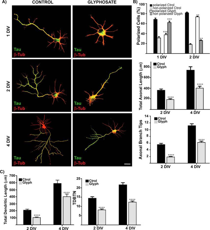
Figure 1 Sub-lethal doses (4mg/ml) of glyphosate induce abnormal morphology of embryonic rat neurones and delayed differentiation; (A) Confocal microscope images of immuno-labelled neurones with the axonal marker Tau1 (green) and β-tubulin (red) following 1, 2 and 4 days in vitro (DIV) culture; (B) Number of polarised cells, total axon length, and total axonal branch tips in glyphosate treated cells following 2, 4 DIV; (C) Total dendritic length and total dendritic branching tip number (TDBTN) in glyphosate-treated neurones following 2 and 4 DIV
Analysis of growth cones (situated on the very tips of developing dendrites and axons that seeks synaptic targets with other neurones) showed that glyphosate caused a significant reduction in their size. Staining for markers of growth cones of minor processes as well as prospective neurones revealed a 70 % and 54 % reduction respectively. Furthermore, the organization of actin (a structural protein abundant in neurones) around the growth cones was disrupted. Neuronal polarisation depends on the organisation of the actin cytoskeleton. Application of a chemical that destabilises actin, cytochalasin D, to neurones was able to abolish the effects of glyphosate, showing that glyphosate neurotoxicity works via disrupting actin cytoskeleton organisation and stability.
With the role of Wnt in the development of neural circuitry well known, the researchers went on to see if any Wnt pathways were dysregulated by glyphosate treatment. First they looked at expression levels of different Wnt proteins. They found that glyphosate treatment reduced the mRNA expression (by approximately 25 %) of one Wnt gene, Wnt5a, after 24 hours of 4 mg/ml glyphosate treatment. After 48 hours, there was also a significant reduction in Wnt5a protein levels. Wnt5 - previously been shown to be involved in axonal branching and growth, axon guidance and neurite development, as well as actin organisation - was disrupted following glyphosate treatment [5-8].
To confirm the involvement of Wnt5a in glyphosate-induced neurotoxicity, recombinant Wnt5a proteins were added to neuronal cultures along with glyphosate; this resulted in neurones with similar morphology to controls (see Figure 2). After 2 days of glyphosate treatment alongside recombinant Wnt5a expression, neurones showed normal axon branching and lengths as well as normal dendritic branching complexity.
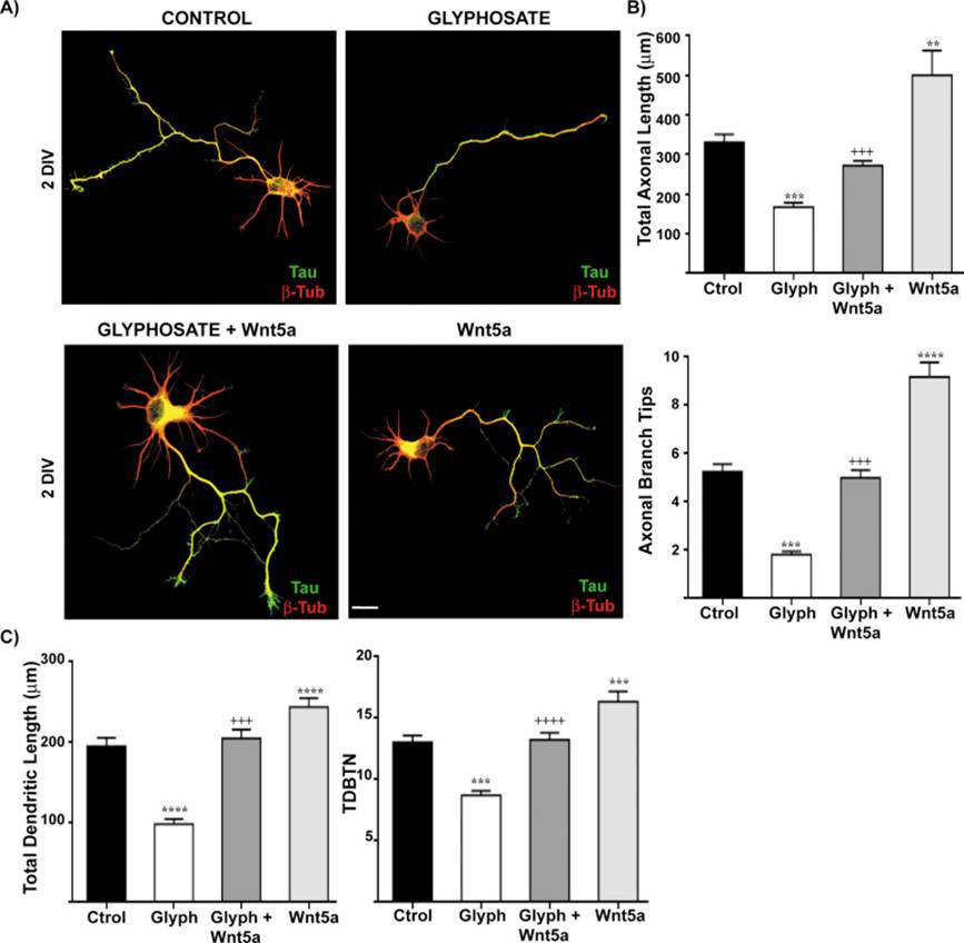
Figure 2 Glyphosate neurotoxicity rescued by addition of Wnt5a protein to neuronal cells; (A) Confocal microscope images of neurones treated with glyphosate and recombinant Wnt5 following 2 days in vitro (DIV) culture; (B) Axonal length and number of axonal branch tips in cells treated with glyphosate with and without Wnt5a; (C) Dendritic length and total dendritic branching tip number (TDBTN) in cells treated with glyphosate with and without Wnt5a
Wnt proteins can signal through different pathways, referred to as the canonical and non-canonical pathways. The canonical Wnt pathway causes the accumulation of β-catenin in cells that act as a transcriptional activator. There are two non-canonical pathways: the non-canonical planar cell polarity pathway and the non-canonical Wnt/calcium dependent pathway. The latter helps regulate calcium release from the endoplasmic reticulum in order to control intracellular calcium levels. Increased calcium levels leads to activation of signalling molecules such as Ca2+/calmodulin-dependent protein kinase II (CaMKII), which is involved in many pathways including those important for learning and memory, development of neural circuits and synaptic plasticity, calcium homeostasis, and reuptake in cardiomyocyte heart cells among other things. The researchers found that following 24 hours of glyphosate exposure, cultured hippocampal neurones showed reduced levels of phosphorylated CaMKII levels by 25 %. The canonical pathways were not altered by glyphosate treatment. Knocking down CaMKII expression using RNA interference techniques abolished glyphosate’s neurotoxic effects on the neurones, indicating that Wnt5a requires CaMKII to elicit its effects on axon growth and elongation. Further, knocking down CaMKII in untreated neurones caused abnormalities similar to that seen under glyphosate treatment, suggesting that glyphosate’s effects work via a reduction in CaMKII activity.
Though glyphosate has been shown to affect both the health of people and planet, new research is helping to elucidate the underlying mechanisms of its toxic effects. This work serves to further confirm the dangers of widespread glyphosate use.
Article first published 20/01/16
Comments are now closed for this article